Search Doctor by:
--Select-- Doctor Name Specialty Zip Code

Blog

All About Male Breast Cancer

Breast cancer is usually thought of as a female disease, and with good reason: Male breast cancer actually makes up less than 1% of all cases of breast cancer. It is most common in older men, although it can appear in men at any age. Because most men ignore signs of breast cancer, such as a lump, male breast cancer is most often detected at an advanced stage, past the point where it can be treated easily.

Chemotherapy and Mitosis

Risk factors for male breast cancer include radiation exposure, a family history of breast cancer, a genetic predisposition to breast cancer (e.g., BRCA1 or BRCA2 mutation), and high levels of the hormone estrogen (usually linked to a disease such as liver disease or Klinefelter syndrome).

Symptoms

The most common sign of male breast cancer is a discernable lump in the breast area, usually just below the nipple. Other symptoms may include skin changes near the nipple, discharge from the nipple that is bloody or opaque, or pain in nearby bones (caused by cancer that has spread beyond the breast). Advanced male breast cancer can also cause general feeling of malaise, weakness, and weight loss.
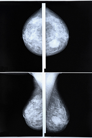
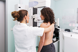
Diagnostic Tests

If your doctor is concerned that you may have male breast cancer, he will order several tests to make a diagnosis. This is usually followed by a clinical breast exam, in which the doctor or professional feels the breast tissue for lumps or other abnormalities. The doctor may then order an ultrasound or an MRI of the breast area in order to see more clearly what the breast tissue looks like and to make an accurate diagnosis.

If the doctor finds a lump, a biopsy may be necessary to determine whether the tissue is cancerous. A biopsy removes some of the tissue, either through using a wide needle or by actually cutting a lump of tissue away from the surrounding area.

Prognosis

If you have received a diagnosis of male breast cancer, your doctor will make decisions about your treatment based on the type of breast cancer, the stage that the cancer has reached, the estrogen-receptor and progesterone-receptor levels in the tissue, your age, and your overall health. In general, the prognosis for male breast cancer is about the same as a female breast cancer prognosis.

Types of Male Breast Cancer

The most common type of male breast cancer diagnosis is infiltrating ductal carcinoma, which originates in the breast ducts and spreads to the surrounding tissue. In contrast, males very rarely get lobular cancers, or cancers that originate in the milk glands, since male breast tissue does not usually contain milk glands. Other common types of male breast cancer include ductal carcinoma in situ (cancer that originates in the milk ducts but has not spread), cystosarcoma phylloides (cancer that originates in the connective tissue around the ducts), and Paget’s disease of the breast (cancer that originates near the nipple).

Keywords: male breast cancer, types of male breast cancer, breast cancer, diagnosis

How Chemotherapy Works

Chemotherapy has become a commonly used term and most people know that it helps to treat cancer and prevent a cancerous tumor from growing. But many people have no idea how chemotherapy works to keep cancer at bay. In order to understand how chemotherapy works, you’ll need to understand the basics about cell division.

Chemotherapy and Mitosis

Chemotherapy and Mitosis

All cells undergo a process called mitosis, or cell division. The cells in fetuses and infants are undergoing mitosis at a very fast rate, whereas most of the cells in adults only undergo mitosis when they need to repair damage. Cancer cells, on the other hand, divide much more often than normal cells. In fact, they undergo mitosis so often that they eventually grow large enough to form a lump, or tumor.

Chemotherapy actually targets cell cells that are in the process of dividing, often killing them. Because cancer cells undergo cell division much more often than normal cells, chemotherapy usually ends up killing cancer cells while leaving the rest of the body’s cells unharmed.

Side Effects of Chemotherapy

There are some non-cancerous cells, however, that divide extremely quickly. For example, hair cells are constantly replenishing themselves and growing. That’s why chemotherapy often causes hair to fall out; the chemotherapy targets the constantly growing hair follicles and kills them, as well as the cancer cells that it is intended to target.

Now that you understand how chemotherapy works, you can easily understand other side effects of chemotherapy. Other cells that undergo cell division regularly include bone marrow cells, skin cells, and cells in the lining of the digestive tract. That means, for example, that the chemotherapy may kill healthy bone marrow cells along with cancer cells. Since bone marrow produces white blood cells that are important to the body’s immune system, chemotherapy treatment may kill off some of the bone marrow cells, necessitating a bone marrow transplant. Since cells that line the digestive tract multiply on a regular basis, they also may be targeted by chemotherapy agents. This can lead to intense nausea and problems with the digestive system in chemotherapy patients.

Why Use Chemotherapy

Although chemotherapy treatment cannot always completely destroy all of the cancer cells, it can sometimes help when combined with other treatments options. For example, a patient with breast cancer may require surgery to remove the cancerous tumor, but chemotherapy can help to reduce the likelihood of the cancer returning. Even for some cancers that are incurable, an oncologist may suggest chemotherapy in order to shrink the tumor, relieve the symptoms of cancer, or prolong the patient’s life by keeping the cancer under control for as long as possible.

In some cases, chemotherapy drugs can help put a patient into complete remission, in which case the cancer cannot be detected at all by using scans, blood tests, or other testing methods. This does not mean that the cancer is gone for good, but it does mean that the cancer is no longer harmful for the time being. In other cases, the goal is for the patient to achieve partial remission, in which the cancerous tumor has shrunk or stopped growing. Sometimes this can enable other treatments to help, such as radiation or surgery.

Keywords: chemotherapy, mitosis, cell division, how chemotherapy works

Surgical Options for Breast Cancer Treatment

Most women with breast cancer will require some sort of surgery as part of their treatment. Surgical options for breast cancer can remove the tumor, the affected breast, the other breast, or the lymph nodes, depending on the spread of the cancer.

Lumpectomy

Lumpectomy

The main surgical options for breast cancer used to remove the tumor are a lumpectomy and a mastectomy. A lumpectomy is the removal of only the tumor itself, along with some of the surrounding tissue, whereas a mastectomy is the removal of the entire breast. This procedure is also called “breast conserving surgery,” “partial mastectomy” or “quadrantectomy.”

A cancer patient who undergoes a lumpectomy usually requires radiation afterwards, and may require chemotherapy as well. In order to ensure that all of the cancer is gone, the surgeon will examine the edges of the removed tissue, called margins. If the patient has clear, or negative, margins, the patient can continue on to the next step of treatment. If the patient has positive margins, the surgeon will need to repeat the surgery to remove more tissue in a surgery called re-excision.

Depending on the amount of breast tissue removed, a lumpectomy may leave the breast looking different than it did originally. A patient may choose to undergo reconstructive surgery either during or after the lumpectomy.

Mastectomy

Mastectomy

There are several different types of mastectomies, or breast removal surgical options for breast cancer. The standard type of mastectomy for treating breast cancer is a simple mastectomy, in which one or both breasts are removed entirely, including the nipple and all breast tissue. In a skin-sparing mastectomy, most of the skin over the breast is left intact, although the nipple and areola are removed. This is ideal for those who intend to reconstruct the breast soon after the surgery. In a nipple-sparing mastectomy, the nipple and areola are left intact. This is only an option for women who have a very small cancerous area far from the nipple. Because some tissue is left in the nipple area, women who have a nipple-sparing mastectomy are at a slightly higher risk of a cancer recurrence and those who had a simple or skin-sparing mastectomy.

Lymph Node Surgery

Lymph Node Surgery

There are two main types of surgery that check the lymph nodes to determine whether the cancer has spread to them. In an axillary lymph node dissection (ALND), ten to forty lymph nodes are surgically removed and examined for signs of cancer. Although an ALND is a relatively safe procedure, it does have some side effects. Therefore, surgeons often opt to begin with a sentinel lymph node biopsy (SLNB), in which the surgeon uses a radioactive dye to find the sentinel lymph node, or the first lymph node to which a tumor would likely spread. The surgeon would then remove that lymph node surgically and check it thoroughly for cancer. There are often two or three sentinel lymph nodes, in which case all of them would be removed during this procedure.

Reconstructive Surgery

Reconstructive Surgery

After a mastectomy or some lumpectomies, the patient may decide to rebuild the breast for aesthetic purposes. Options for reconstructive surgery include immediate or delayed reconstruction. The patient could also choose from several materials that could fill the new breasts, such as saline, silicone gel, cohesive gel (colloquially called “gummy bears”), or even the patient’s own tissue from elsewhere on the body. Reconstructive surgery does carry risks, however, and may need to be repeated over the course of the patient’s life.

Keywords: mastectomy, lumpectomy, reconstructive surgery, surgical options for breast cancer

Breast Cancer Risk Factors

In order to make mature decisions about breast cancer screenings, it is important to understand the risk factors of breast cancer. Keep in mind, however, that having a risk of breast cancer does not mean that you’re destined to develop it; most women with one or two breast cancer risk factors will never develop breast cancer at all.

Gender, Age, and Race/Ethnicity

Gender, Age, and Race/Ethnicity

Although men can get breast cancer, women are about a hundred times more likely to get it than men are. Besides the fact that women have more breast tissue, they also produce more of the hormones estrogen and progesterone, which can promote the growth of cancer. Age is a risk factor for breast cancer as well, with about two thirds of breast cancer patients being above the age of 54. White women are also more likely to develop breast cancer than most other nationalities, although cancer in African American women is more likely to be aggressive, caught at a later stage, and diagnosed at an earlier age.

Family and Personal History

Family and Personal History

If you have even one close relative that has been diagnosed with breast cancer, you have twice the risk of developing breast cancer than you would otherwise. Furthermore, if you have personally been diagnosed with breast cancer in the past, you are 3 to 4 times more likely to develop additional breast cancer, either in the other breast or in a different area of the same breast. (This is different from the risk of recurrence, which is the risk of the original breast cancer itself returning.)

Genetics and Hereditary Cancer

Genetics and Hereditary Cancer

Between 5% and 10% of breast cancer cases are believed to be hereditary cancer. The most common genetic cause of breast cancer is a mutation in the BRCA1 or BRCA2 genes. In normal cells, these genes code for the production of proteins that prevent cells from growing abnormally. In other words, they indirectly prevent cells from becoming cancerous. A mutated copy of these genes, however, does not work as well, which is why women and men who inherit these mutations are at a higher risk of developing breast cancer (as well as some other cancers). This type of hereditary cancer often occurs in younger women and is more likely to affect both breasts. Other gene mutations can also put you at a higher risk of developing hereditary cancer, although these are much rarer than the BRCA mutations.

Other Breast Conditions

Other Breast Conditions

Some other issues with the breasts or breast tissue can also put you at a higher risk for breast cancer. For example, women with dense breast tissue may have up to twice the likelihood of developing breast cancer; unfortunately, dense tissue can also make mammograms less accurate. Some benign breast conditions – although not all – can also act as risk factors for breast cancer. Some, such as ductal hyperplasia and fibroadenoma, only increase the risk of developing breast cancer slightly. Atypical ductal hyperplasia (ADH) and atypical lobular hyperplasia (ALH), on the other hand, can raise your risk of developing breast cancer up to five times higher than normal.

Other Risk Factors

Other Risk Factors

Women who began menstruating early or who went through menopause later, and therefore experienced more menstrual cycles and the resulting hormones, are at a slightly higher risk of developing breast cancer. Women who have had radiation treatment to the breast area (e.g., lymphoma), on the other hand, have a significantly higher risk, especially if the radiation treatment occurred during childhood or adolescence. In addition, women who took the drug diethylstilbestrol (DES), which was given from the 1940s to the 1960s to prevent miscarriage, have a slightly higher risk of developing breast cancer.

Keywords: breast cancer risk factors, risk of developing breast cancer, breast cancer, hereditary cancer

How to Do a Breast Exam

The National Breast Cancer Foundation encourages women of all ages to perform a breast self-exam at least once a month in order to find early signs of breast cancer. A self-exam involves feeling the tissue of your breast and the surrounding area in order to identify any changes that could signify a cancerous lump or thickening. Even women over age 40 who receive an annual mammogram, which can detect breast cancer before you are able to feel a lump, should perform a self-exam in order to familiarize themselves with the “normal” look and feel of their breasts so that they can identify any changes. To keep track of your monthly self-exam, you may want to take a calendar and fill it in on each month, do it every month on the day you get your period, or put a monthly alarm on your phone.

There are three main components of a breast self-exam. The first should be performed in front of a mirror, the second in the shower, and the third while lying down. This will ensure that you have increased your risk of identifying any tissue changes from as many angles as possible. Keep in mind, however, that pregnancy and nursing can change the feel of your breasts due to modifications in the milk ducts.

In Front of a Mirror

In Front of a Mirror

While standing undressed from the waist up in front of a mirror, look carefully at your breasts. Check for any changes in shape, size, or position, as well as for any changes to the skin itself, such as discoloration, dimpling, or tightness, any of which could be a sign of breast cancer. Repeat this process with your hands pressing on your hips so that the muscles beneath your breasts tighten, bending forward with your shoulders pushing forward, and clasping your hands behind your head with your elbows pressing backwards. Make sure to lift your breasts to check the skin on the underside and to gently pull on your areola to check for any abnormal discharge.

In the Shower

In the Shower

Besides for visually examining the breasts, you’ll want to feel the breast tissue to identify any changes. The easiest location to do this is in the shower so that your fingers will be able to move smoothly over your breasts. Make sure to feel both underarms, above and below your collarbone, and the entire breast from bra line to collarbone, for unexpected lumps or thickenings. It is easiest to do this with one arm raised behind your head and the other hand feeling with the flat surface of your fingers.

Lying Down

Lying Down

You may want to moisten your fingers with body lotion to complete this last component of a breast exam. To spread out the breast tissue, lie down with your right shoulder on a small pillow and your right arm behind your head. Then use the flat fingertips of your left hand to feel in small circular motions around the entire area of the breast. Move slowly, taking note of any bumps or uneven sections of breast tissue. Pay special attention to the nipple area, making sure that there are no lumps or other signs of breast cancer beneath it and that you gentle pressure can easily push the nipple slightly inward.

Keywords: breast self exam, breast cancer, breasts, breast tissue

Managing Your Risk of Developing Breast Cancer

If you’re at a high risk for breast cancer, it can seem like a death sentence. Luckily, you can reduce your chances of developing breast cancer, or increase your risk of catching it early, by understanding the research behind breast cancer development and breast cancer screenings.

Lifestyle Changes

Lifestyle Changes

Lifestyle changes can help you reduce your chances of developing breast cancer. For example, limiting alcohol, smoking, and exposure to radiation or environmental pollution has been shown to lower breast cancer risk. To further reduce your risk, stick to a healthy lifestyle to ward off the risk factor of obesity, making sure to include plenty of physical activity in your day.

Hormonal Changes

Hormonal Changes

Having children young has been found to reduce your risk of developing breast cancer. Breast feeding has been proven protective as well, with this protection increasing based on the amount of time you have breastfed. Hormone replacement therapy (HRT) should be avoided as much as possible in women who are at high risk for breast cancer. Research shows that women who stop HRT have a decreased risk of developing breast cancer later in life.

Breast Cancer Screenings

Breast Cancer Screenings

If you are at a high risk for breast cancer, you can manage your risk by taking part in various breast cancer screenings and tests. The most basic breast cancer screenings are the breast self-exam and the clinical breast exam. A breast self-exam involves checking your own breast tissue, including the tissue under your arm and around your nipple, for lumps or irregularities. Your doctor can also perform a clinical breast exam, which follows the same protocol.

Beyond the basic breast exam, the most common screening test for breast cancer is a mammogram, which x-rays the breast tissue to find potentially cancerous masses. The American Breast Cancer Society recommends that all women begin getting an annual mammogram starting at age 40, but women with a higher risk of developing breast cancer (e.g., BRCA mutations, strong family history) may begin receiving mammograms years earlier. Mammograms can identify lumps in breast tissue up to two years before they can be felt by a breast self-exam.

If you are at high risk for breast cancer, your doctor may also recommend a magnetic resonance imaging test (MRI). This test uses a strong magnetic field and pulses of radio frequencies to produce an image of the breast tissue. An MRI is not a replacement for a mammogram, but it can be used in conjunction with a mammogram in order to identify masses that the mammogram missed or evaluate a mass that was hard to see on a mammogram.

Keywords: breast cancer, breast cancer screenings, risk of developing breast cancer, high risk for breast cancer

Types of Breast Cancer

There are several different ways to classify types of breast cancer, and the way that a case of cancer is described can incorporate several of these classifications. For example, a triple-negative case of invasive lobular carcinoma includes classifications for the hormone receptivity of the cancer cells (“triple-negative”), the invasiveness of the cancer (“invasive”), the location of the cells (“lobular”), and the origin of the cells (“carcinoma”). Understanding the different types of breast cancer can help you make educated choices about your treatment options.

Cancer Origins and Invasiveness

Cancer Origins and Invasiveness

Biopsied tissue is examined under a microscope to determine whether cancer is present, as well as to find out more about the cancer’s origins and invasiveness. Most breast cancers are carcinoma, which means that they develop from epithelial cells. Cancer can also be labeled “sarcoma,” which means that it originated in non-epithelial cells, but this is rare for breast cancer.

Cancer is also categorized based on whether it is invasive, which means that it usually “invades” or spreads outside of the milk lobules or ducts. Breast cancer usually originates in the milk lobules or ducts, but non-invasive breast cancer rarely metastasizes beyond them. The phrase “in situ” means that the cancer is non-invasive or pre-invasive (it has not yet spread to the rest of the tissue).

Cell Location

Cell Location

Breast cancer is also categorized based on where the cancer cells are located within the breast. The cancer cells in ductal carcinoma are located in the milk ducts, the cancer cells in lobular carcinoma are located in the milk-producing glands, which are called lobes. Lobular carcinoma is often more difficult to detect by mammogram than ductal carcinoma.

Hormone Receptors

Hormone Receptors

You can also classify a breast cancer based on which hormone receptors it has. Two hormones often promote the growth of breast cancer cells: estrogen and progesterone. Normal breast cells contain hormone receptors that attach to both of these hormones, and some breast cancer cells do as well. Cells that contain at least one of these hormone receptors are called “receptor positive.”

In addition, about 1 in every five breast cancers have increased levels of a protein called HER2/neu that promotes cell growth. These cancers are called “HER2 positive,” and can be treated with drugs that target HER2.

Doctors examine breast cancer cells to determine whether they have receptors for estrogen, progesterone, or HER2/neu. Based on their findings, they decide on the type of treatment that will be most successful. If a cancer has none of these receptors, it is said to be “triple negative,” and cannot be treated with therapies that target HER2, estrogen, or progesterone. Triple negative cancer has fewer treatment options, although it can still be treated with chemotherapy. Triple negative cancer also tends to grow more quickly than other cancers.

Less Common Types of Breast Cancer

Other, less common types of breast cancer include the following:

  • Inflammatory breast cancer, which leaves the breast skin looking red and warm, and often with the texture of an orange peel
  • Paget disease of the nipple, which leaves the skin of the nipple and areola red or crusted, possibly secreting blood or other liquid
  • Phyllodes tumor, which develops in the connective tissue, or stroma, of the breast, rather than in the milk ducts or lobules
  • Angiocarcinoma, which originates in the cells lining blood vessels or lymph vessels.

Keywords: breast cancer, hormone receptors, types of breast cancer, triple negative

Lumpectomy Vs. Mastectomy

Making the decision between a lumpectomy and a mastectomy can be difficult. It’s a decision that is often left up to the patient, rather than the doctor. Understanding the advantages and disadvantages of each choice can help you make a more informed decision about the treatment option you choose.

A lumpectomy, also called “breast conserving surgery,” is the removal of the cancerous lump in the breast, as well as a margin of tissue surrounding the cancer. A lumpectomy usually also requires radiation therapy, even if the cancer is limited to the breast tissue. A mastectomy, on the other hand, is the removal of one or both breasts, sometimes including the lymph nodes under the corresponding armpit. These two procedures have an almost identical success rate in terms of survival, but a lumpectomy has a slightly higher chance of the breast cancer recurring.

Advantages of a Lumpectomy

Advantages of a Lumpectomy

The main advantage of a lumpectomy is that the breast is conserved as much as possible. It is also a more minor surgery than a mastectomy, with a shorter and easier recovery time and fewer potential side effects. You can usually go home later in the day after a lumpectomy, rather than staying overnight in the hospital, as with a mastectomy.

Disadvantages of a Lumpectomy

Disadvantages of a Lumpectomy

Although the idea of keeping your beasts virtually intact can make the prospect of a lumpectomy more inviting than that of a mastectomy, a lumpectomy does have several potential disadvantages. First of all, a lumpectomy is usually followed by five to seven weeks of radiation therapy, to ensure that the cancer has been completely removed. The radiation therapy can affect the timing of any reconstruction, as well as your reconstruction options, depending on the extent of the radiation therapy needed.

In addition, a lumpectomy does have a higher chance of a local recurrence of breast cancer than a mastectomy. If the same breast experiences a recurrence of breast cancer, additional radiation therapy is no longer an option, which means that you would have to undergo a mastectomy anyway.

Factors to Consider In order to decide which procedure is best for your specific case, it is important to ask yourself some important questions. For example, how important is it to you to keep your original breast? Breast reconstruction is an option for those who opt for a mastectomy, but the breasts will not experience sensation and may require multiple additional surgeries in order to ensure that they are aesthetically pleasing. Also, you’ll have to consider the size of the cancer that would be removed by a lumpectomy. If the lump is very large, a lumpectomy may leave you with a distorted breast that can be very difficult to repair to your approval. In addition, your level of anxiety about recurring breast cancer should be taken into account, since a mastectomy radically lowers your risk of recurring cancer.

Keywords: mastectomy, breast cancer, lumpectomy, radiation therapy

Recovering from a Mastectomy

Whether you’ve had breast cancer or are known to have a predisposition to breast cancer, your doctor may recommend a mastectomy. A mastectomy, or removal of one or both breasts, is a major surgical procedure that requires plenty of recovery time. Whether your mastectomy was due to breast cancer or a predisposition to breast cancer, keep in mind during the recovery that the surgery may have saved your life, and that the pain and discomfort will end and life will go on. In the meantime, you can make the recovery period easier by following your doctor’s instructions and knowing what to expect.

Pain Management

Pain Management

You’ll want to fill the prescriptions for your pain medicines as soon as possible, and take them before the pain hits. Because it’s easy to lose track of time after surgery, use a notebook to write down the times and doses of pain medicines that you’ve taken. Your doctor will also give you laxatives, since you may experience constipation after the surgery.

Drains, Stitches, and Staples

Sometimes, the drains will be removed before you leave the hospital. In many cases, however, you will go home with multiple drains still in for a week or two following the surgery. If so, you’ll need to empty the bulb multiple times a day and keep track of how much fluid you are emptying out.

Your incision will be closed with either stitches or staples, probably covered by a bandage. Stitches usually dissolve over time, and your surgeon can easily remove a stitch that pokes out of the incision. Staples need to be removed by the doctor, which would usually happen during your first post-op visit. Do not remove any bandages or stitches unless you have your doctor’s instruction to do so.

Rest and Recovery

Rest and Recovery

Perhaps the most important piece of advice you can get about recovery from a mastectomy is the importance of resting and taking it easy. Fatigue is completely normal after a mastectomy, and you should avoid doing anything more strenuous than walking – including housework, if possible – until the drains have been removed. Depending on your recovery time, you will probably be ready to go to work between three and six weeks after the surgery.

If you have had lymph nodes removed, it may be difficult or uncomfortable for you to move your arm. Although you should not specifically avoid using your arm, go slowly; even pulling open a drawer can cause you pain during the first week if you’re not careful. Try to put objects that you will need often at an easy height, and keep in mind that you may need help with basic actions such as pulling a coat on or opening a child-safe pill cap. You should also avoid exercising your arms strenuously until after you have seen your doctor at your first post-operative visit and avoid heavy lifting for six weeks following the surgery. In time, you’ll be given exercises to keep your muscles flexible and to improve your range of motion.

Make sure to take care of yourself after your mastectomy, taking any offers of help from family or friends seriously, and accepting them whenever possible. The more you rest, the easier your recovery will be. Make sure you’re eating nutritious food and drinking plenty of water as well.

Sleeping

Sleeping

Although you’re supposed to be getting a lot of rest, many people find it difficult to sleep after a mastectomy. You will need to sleep on your back for three or four weeks, which can be difficult if you’re used to sleeping on your stomach or side. Not only that, but laying your arms by your side can pull at the surgical site, causing discomfort or pain while you sleep. You also may wake up several times a night due to muscle spasms or the need to move around, especially if you had expanders inserted during the surgery.

Again, keep in mind that the discomfort is temporary, and that the surgery can virtually eliminate the risk of recurring breast cancer.

Keywords: mastectomy, breast cancer, surgery, recovery

Mammograms: What to Expect

A screening mammogram is a specialized X-ray that takes images of male or female breasts in order to identify potential sources of cancer. The age that you begin going for mammograms differs based on your family history of breast cancer, genetic factors that can make you predisposed to cancer (such as the BRCA genes), or a personal history of other cancers. Because it is normal to be nervous before your first mammogram, you should take the time to educate yourself about what to expect. Mammograms can be uncomfortable, or even painful, but are necessary in order to ensure that you don’t currently have an early stage of breast cancer.

Preparing for the Procedure

Preparing for the Procedure

Make sure to avoid dresses or other one-piece clothing on the day of your mammogram. If you are sensitive to pain, you may want to take a painkiller before the procedure. Soon after you arrive, you will be escorted to a small dressing room where you can remove all clothing from the waist up, as well as any jewelry or other accessories. The attendant will provide you with a gown that ties in the front, and may allow you to place your clothes and other belongings in a secure cabinet.

Taking the X-ray

Taking the X-ray

A technician will take you into a private room with a large X-ray machine used specifically for mammograms. In order to get an accurate picture, the technician will need to manually place your breast onto a platform and manipulate it so that it is positioned correctly. The technician will also ask you to move the rest of your body so that the mammogram can focus on the breast tissue without any obstructions.

During the actual X-ray, your breast will then be sandwiched between two plates so that the X-rays can penetrate the tissue, as well as so that breast tissue remains completely still. This can cause some discomfort, and even pain, but lasts for only a few seconds at a time. During this time, you should stand as still as possible and hold your breath when the technician asks you to.

After a Mammogram

It is extremely common to need to retake an X-ray due to some concern that the technician will find while reviewing the images. Most of the time, these concerns do not end up being cancer, so expect to be called back from the waiting room a second time. If you’re one of those lucky ones whose pictures come out perfectly the first time, you’ll be happily surprised.

You will not receive the results of a mammogram the day of the test. In the US, the facility legally needs to send you the results within 30 days, but they usually arrive even sooner.

Keywords: mammogram, breasts, cancer, X-ray